Hi, I’m Aneesh. I wanted my own H10’s raw ECG without a subscription, so I built an app and I’m giving it away. Everything is free and stays free. No ads, no account.
Feature
What you get
Live ECG
raw, 130 Hz, zoom
Morning check
3 min: resting HRV, heart rate, breathing vs your own usual range
Waveform
QRS, QT, QTc, PR, J point, ST, T waves
Breathing
from the strap’s motion sensor + ECG
Threshold test
DFA α1 + breathing rate
Export
CSV with every sample, or PDF. Free.
Accuracy
Result
Tested on
Beats
99.6 % found
MIT-BIH
RMSSD, SDNN, pNN
identical to NeuroKit2
MIT-BIH
DFA α1
±0.001 vs NeuroKit2
MIT-BIH
Breathing
±0.1/min paced, 97 % of minutes within 2/min
metronome, BIDMC
QRS · PR · QT
80 · 78 · 67 % within 20 ms of cardiologists
QT Database, LUDB
ST · T height
88 · 70 % within 25 µV
QT Database, LUDB
J point
41 % within 25 µV (two cardiologists agree on 45 %)
QT Database
Threshold HR
not yet tested against a lab
method after Rogers 2023, simplified; I’m porting the exact method
All at the H10’s 130 Hz. Full table with limits in the app: Settings → About → Accuracy.
EDF/EDF+: yes, I’ll add it. The ECG (μV, 130 Hz) and the accelerometer go in as signals, markers and connection gaps as EDF+ annotations, so it opens directly in EDFbrowser. It’s on the list for the next release.
Ectopic beats: I detect beats from the raw ECG myself, not from the strap’s RR intervals. Every beat is then classified with the method of Lipponen & Tarvainen 2019: normal, ectopic, missed, extra, long/short or artefact, with thresholds that follow the surrounding 91 beats. RMSSD, SDNN and pNN50 use only intervals between two normal beats, so a flagged beat is left out, not patched. The app shows how many were left out. One limit, which is on the Accuracy page: single ectopic beats are caught well (28 of 30 on MIT-BIH record 100), but when they come very often most pass as normal. I’m working on flagging those recordings.
mV on the live plot: To be honest, I had it in the very first internal version, then removed it. The trace drifts when the strap moves, so an absolute axis would be wrong. It can be done the way ECG paper does- a fixed grid in mV per division with a 1 mV calibration bar, plus the R-wave height of the last beats as a number can be done, but I cannot guarantee it in the very next version.
Hi pepe, version 0.9.0 is out on Google Play, and two of your three points are in it.
EDF+ export. Every recording now has “Export EDF+” in its Export menu. The file holds the ECG at 130 Hz, stored as recorded at 1 µV per bit, and the accelerometer’s X, Y and Z at 25 Hz in mg. Annotations mark the recording start, each stage of a threshold test, every connection gap with its length, and the end with its reason. The header is anonymous. I checked the files with EDFbrowser’s own compatibility check, and with pyedflib and MNE. Two things to know: a gap of up to 5 s is joined and marked with an annotation, while a longer gap starts a new file; and EDF stores whole seconds, so the last fraction of a second is only in the CSV.
How much data that is: 130 ECG samples and 75 accelerometer values per second, so about 468,000 ECG samples per hour. The CSV export still has every sample with its timestamp.
Voltage on the live ECG. Done the ECG-paper way I described: a fixed grid where each square is 0.2, 0.5 or 1 mV, plus a 1 mV calibration pulse. The R-wave height as a live number is not in yet.
Ectopic and irregular beats. The handling is as I wrote before. New: when the time between beats was irregular for 5 minutes or more, the reading shows a “Time between beats” chart with those stretches marked, and HRV is then calculated from the regular part only.
Also new: a “Heart rate and breathing” mode for long recordings (up to 24 hours, no ECG; the breathing rate comes from the strap’s motion sensor and is marked experimental), an “Average heartbeat” picture for each reading, and backup and restore.
If you try the EDF+ export in your EDFbrowser setup, I would be glad to hear whether it opens as you expect and what is missing.
Hi Aneesh,
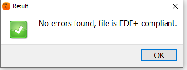
I briefly tested the 0.9.0 EDF+ export in EDFbrowser and ran its built-in compatibility check—it passed with “No errors found, file is EDF+ compliant”
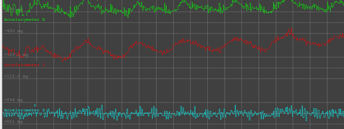
At rest the 130 Hz ECG and 25 Hz 3-axis accelerometer channels (X, Y, Z) all load with accurate physical scaling, clearly showing the respiratory oscillations and mechanical heartbeat spikes. Everything works as expected!
Thanks pepe, good to see EDFbrowser’s own check pass on your side.
You spotted the breathing wave in the accelerometer. The new Heart rate and breathing mode is built on that: two different estimators run on the last 60 s, and a rate is shown only when you are still and both agree within 2 breaths/min.
It works sitting or lying, and breaks with slight movement. In daily-life data it shows a value in only about 14 % of the still windows.
Any ideas how to keep the breathing signal when the torso moves a little? You know the Tymewear data, so I am curious how that behaves.
It is all written up: paper (PDF). Results are on page 8, the method on pages 21 and 22, and the step-by-step algorithm from page 34.
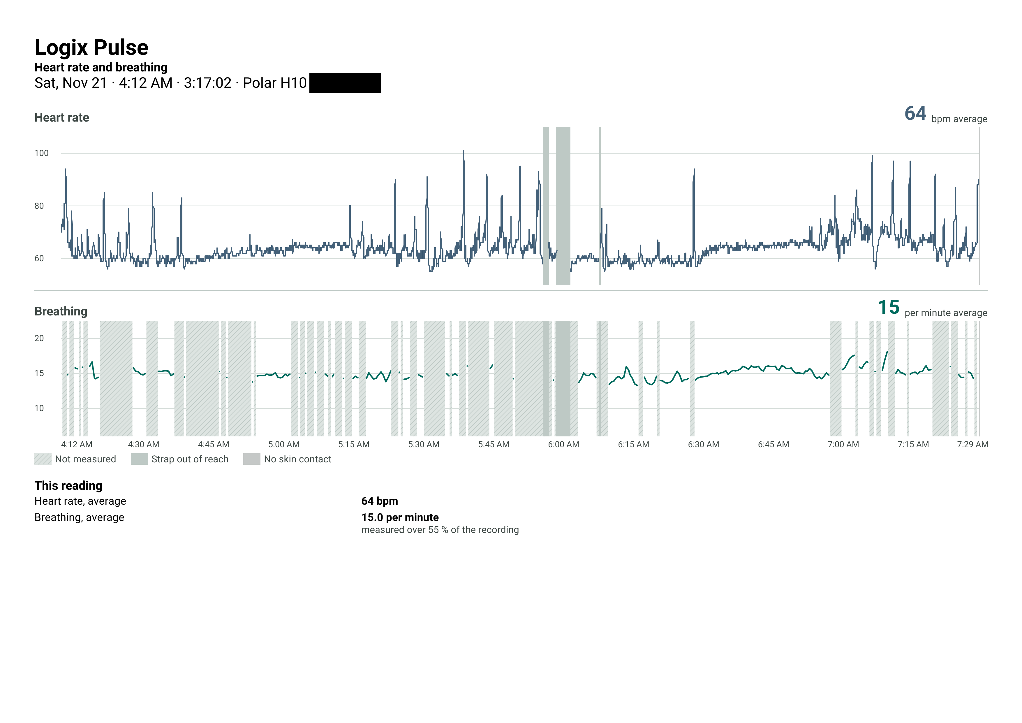
I’m also attaching an Export for the Logix Pulse HR and Breathing Mode that I recorded this evening during my workout. I only got breathing measurements 5% of the time. I think its still better that nothing, and I’m not sure if any other app is even doing this. But I want to improve it.
Thanks for sharing the paper—the level of detail in M13 and Supplementary Methods S6 makes it super clear why coverage drops to ~14% in still daily-life windows and ~5% during a workout.
Why Tymewear behaves differently Tymewear doesn’t rely on an accelerometer for breathing—its strap embeds a capacitive stretch sensor around the chest that measures circumferential ribcage expansion directly. Because it measures strain rather than inertial acceleration, torso tilt (gravity vector rotation) and cadence/road vibration don’t swamp the respiratory waveform the way they do on a single-point 3-axis accelerometer. Even in accelerometer respiratory papers like Schipper et al. (2021) and Hostrup et al. (2025), accuracy and coverage depend heavily on posture/gravity projection and motion gating, with tight error limits trading off against lower coverage.
How different activity types affect the breathing signal
Gravity and chest movement are relatively steady, so the main challenge is low signal amplitude, shallow breaths, or slow breathing rates where spectral peaks or breath-duration variability V can get penalized.
Slight posture shifts or leans can still tilt the gravity vector and pull the projection away from pure ribcage motion.
Torso motion is often smoother and more continuous than running, with fewer sharp vertical shocks, though pedal cadence and upper-body bracing can still create rhythmic acceleration and posture shifts.
Because the 15 mg window RMS limit and 60 mg single-sample movement gate (which spoils a 4 Hz sample/30-s frame and triggers 10 s of fast settings) are strict, even steady riding easily gets gated out.
Walking:
Introduces periodic step cadence and vertical/anteroposterior oscillations that can overlap with or leak into the respiratory band and shift principal axes away from chest expansion.
Occasional arm swings, turns, or posture changes also trip movement checks and add variability.
Running:
This is the toughest case by far. Footstrikes create sharp vertical shocks and high-amplitude acceleration bursts, while breathing becomes faster, deeper, and more variable.
Both the 15 mg window RMS limit and 60 mg per-sample movement check will trip almost continuously.
Rhythmic bounce and cadence harmonics can pull PCA projections or peak/breath counters off chest expansion—as your limitation notes, rhythmic movement at a breathing frequency can even be read as breathing (such as a 1-mg sine at 45 per minute shown as 45 breaths/min in 21 of 39 windows in simulation), and higher breath-duration variability V can pull quality Q down.
For running specifically, accelerometer-only estimation on a chest pod is probably best treated as mostly gated out or down-weighted rather than forced through heavy footstrike noise.
Ideas to preserve the breathing signal during slight torso movement (M13) Looking at your M13 / S6 pipeline, a few adjustments could recover more windows during still/light movement and smooth cycling without sacrificing your low error rate, while treating running as mostly gated out or down-weighted:
Decouple posture shifts from breathing via faster gravity tracking / high-pass filtering:
In Estimator A, your normal gravity update (alpha_g = 0.03 at 16 Hz) lags behind slow torso leans, letting gravity leak into the horizontal plane h_n. Adding a 0.08–0.1 Hz high-pass filter to the raw axes before projecting onto the horizontal plane (or using a faster complementary/median filter for gravity) prevents slow torso tilts from masquerading as huge breathing swings.
Similarly, softening the 60 mg single-sample movement threshold (which currently invalidates an entire 30-s frame plus a 10-s recovery tail)—for instance, clipping/winsorizing brief movement transients instead of poisoning 40 seconds of data—would stop a single reach or posture adjustment from wiping out two consecutive windows.
Notch or filter cadence harmonics during rhythmic movement (walking/cycling):
During cycling or walking, torso motion has periodic cadence components and harmonics. Even when attenuated by your 0.8 Hz low-pass, strong cadence energy can bleed into the respiratory band and shift the principal component axis toward bounce rather than chest expansion.
Identifying dominant cadence peaks above 1.1 Hz in ||a|| and applying a narrow notch filter—or computing the PCA covariance matrix on signals already band-passed to 0.15–0.75 Hz—helps keep the first eigenvector locked onto ribcage expansion rather than bounce.
SNR-based activity gating instead of a fixed 15 mg hard cutoff:
A fixed 15 mg RMS threshold treats smooth, rhythmic motion (like steady indoor cycling) the same as chaotic movement. Comparing in-band respiratory power (0.15–0.75 Hz) against out-of-band motion power (respiratory SNR) rather than raw RMS ||a|| allows windows with gentle movement to pass as long as the breathing peak remains clearly dominant.
Really impressive work documenting every equation and failure mode openly—especially the H10 0.98 Hz high-pass filter characterization.
References
Prasobhan, A. (2026). How Logix Pulse analyses a Polar H10 recording: every step, the strap’s built-in filter, and what the results can support. LOGIXMOTION.
Schipper, F., van Sloun, R. J. G., Grassi, A., Derkx, R., Overeem, S., & Fonseca, P. (2021). Estimation of respiratory rate and effort from a chest-worn accelerometer using constrained and recursive principal component analysis. Physiological Measurement, 42(4), 045004.
Hostrup, M. C. F., Nielsen, A. S., Sørensen, F. E., Kragballe, J. O., Østergaard, M. U., Korsgaard, E., Schmidt, S. E., & Karbing, D. S. (2025). Accelerometer-based estimation of respiratory rate using principal component analysis and autocorrelation. Physiological Measurement, 46(3), 035005.
Wouldn’t indoor rowing on a static erg (i.e. C2 RowErg) be the toughest case for this? Aside tilting upper body between the 1 and 11 o’clock position, there is considerable acceleration backward during the drive, and a modest acceleration forward during the recovery. People’s body keep moving up and down about a meter. Garmin actually uses the accelerometer for stroke detection on cheaper watches (more expensive ones get this data from the erg directly).
Good point, Jaap. Rowing is probably the worst case. A big, regular movement at 18 to 36 strokes per minute, right inside the breathing range, so the accelerometer sees the stroke and not the breath.
Today the app detects thisusing a fixed “still” limit: RMS of the acceleration magnitude, high-passed at 0.7 Hz, at most 15 mg over the 60 s window. (From my own recordings: under 10 mg sitting or lying still, about 69 mg when changing from sitting to lying.) A rower is far above it, so the app shows “not measured”.
I want to replace that fixed limit. It discards gentle movement where breathing is still readable, yet it is the only thing that keeps your case from going wrong. The plan: check the signal itself for a clear breathing peak that is not the movement rhythm. This will be a hard problem to solve.
But however, I can somewhat use it for sleep tracking. I had a really short and bad sleep the other day, and I was able to detect my breath rate for 55% of the recording. Not yet sure what to do with this. May be sleep quality can be detected better, I’m not sure. But this sleep was definitely not the best
In my ECG analysis of the H10 data during rowing, breathing causes a wave in the R-Wave amplitude. I didn’t persue it further (my goal is detection deviations of regular sinus rhythm), but it seems to be a thing:
I am interested in seeing if RR intervals can be used for breathing, as the rowing app I’m building already captures RR-intervals, but breathing is much more interesting for us (in rowing breathing actually synchronises with the stroke, so a switch from 1 breath per stroke to 1.5 or 2 is extremely telling).
Yes, it is a real effect, and Logix Pulse already uses it on the server: QRS amplitude and RR intervals both go into its breathing estimate (paper, Methods M8, page 16 - 17).
My experience: fine at rest, unreliable quickly. On public daily-life data with a breathing belt, the ECG-only estimate was off by more than 2 breaths/min in 31 to 65 % of the values it showed, even while sitting.
For rowing there is an extra trap: if breathing locks to the stroke, a wave in R amplitude at stroke rate could be the breath or just the movement. A switch to 1.5 breaths per stroke might be the one thing that stands out.
To be clear: that ECG-based estimate is only in the modes that record the ECG (morning check, free recording, threshold test). The long “Heart rate and breathing” mode records no ECG and uses the accelerometer alone (Methods M13).